Pathophysiology of Liver Diseases
| ✅ Paper Type: Free Essay | ✅ Subject: Biology |
| ✅ Wordcount: 1525 words | ✅ Published: 07 May 2018 |
- ONG SUAN LING
Liver Failure
The liver is responsible for regulatory, detoxification, metabolic and synthetic activities. Liver cell known as hepatocyte, is responsible for about 500 or more specific biologic processes [2].
Liver failure occurs when large parts of the liver become damaged and the liver is dysfunction [1]. Liver failure occurs over many years and gradually. However, the acute liver failure is difficult to detect initially and occurs rapidly (as rapid as 48 hours) [1].
Chronic liver failure is caused by excessive alcohol intake, Hepatitis B or C, malnutrition, Hemochromatosis (body absorb and store too much iron) and cirrhosis (irreversible inflammatory disease) [2].

Figure 1 show the stages of liver damage which eventually lead to cirrhosis.
Liver Hepatitis
Hepatitis means inflammation of the liver. Hepatitis B and hepatitis C is the most common hepatitis which can lead to the liver damage caused by their chronic forms. Viral hepatitis is cleared from the body in a period ranging from weeks to months, by the immune system but when it is not as seen in chronic hepatitis, the disease must be managed medically [5].

Hepatitis can also be brought on by excess alcohol consumption or inherited (congenital hepatitis)[5]. When hepatitis left untreated, it can damage the liver over many years, eventually resulting in cirrhosis [5].
Liver Cirrhosis
Cirrhosis is a condition in which normal, healthy hepatocyte are damaged and replaced by nodular and fibrotic tissue. A cirrhosis-damaged liver can cause decreased hepatic function and widespread disruption of many body functions [3].
For cirrhosis, the biliary channels become obstructed and caused portal hypertension. The hypoxic necrosis is developed as a result of blood circulation is shunted away from the liver (due to neovascularisation) [4]

Figure 2 shows the normal appearance of the cells of the liver, compared to cells of cirrhotic livers.
Hepatopulmonary syndrome (HPS)
Hepatopulmonary syndrome is the clinical relationship between hepatic dysfunction and the existence of pulmonary vascular dilatation which can result in a range of arterial oxygenation abnormalities [6].
HPS is defined by the presence of chronic liver disease; abnormal arterial oxygenation or an arterial partial pressure of oxygen in the absence of an alternate cause; and evidence of intrapulmonary vascular dilatations (IPVDs) [7]
Pathophysiology
Hepatopulmonary syndrome occurs mostly in patients who have established cirrhosis and portal hypertension [8]. From a pathophysiological point of view, abnormal intrapulmonary vascular dilatation is linked to portal hypertension, which in itself leads to altered bowel perfusion and an increased rate of enteral translocation of gram-negative bacteria and endotoxin.
This process in turn stimulates the release of vasoactive mediators, which include tumour necrosis factor, haem-oxygenase-derived carbon monoxide, and nitric oxide. The increased production of nitric oxide in the lung plays a central part in the pathogenesis of the hepatopulmonary syndrome [8].
Increased concentrations of exhaled nitric oxide are positively correlated with the increase of alveoloarterial oxygen difference. The constitutive and the inducible isoforms of nitricoxide synthase have been implicated in this process.
In addition, the endothelin system, especially abnormal activation and increased expression of endothelial type B endothelin receptors, is implicated in the pathogenesis of the hepatopulmonary syndrome [8]. In patients who have pulmonary hypertension, endothelin predominantly exerts vasoconstrictive and mitogenic effects due to activation of type A and type B endothelin receptors on pulmonary arterial smooth muscle cells [8]. (Refer to figure 3)

Figure 3 show in presence of portal hypertension, hepatic production occurs of endothelin-1 and expression of endothelial type B receptors, but no type A receptors increase in pulmonary vasculature. Signaling via endothelially expressed endothelin B receptor leads to increase NO production by eNOS, with the overall effect of pulmonary vascular dilatation.
Below is how defective synthesis and metabolism of pulmonary vasoactive substance lead to intrapulmonary Vasodilation.
- Due to the vasodilation and development of shunt, the blood flow is not uniform leading to ventilatation-pefusion mismatch and also restricts the oxygen molecules to reach the centre of the capillaries and hemoglobin in the erythrocyte [9]. (Refer to figure 4)

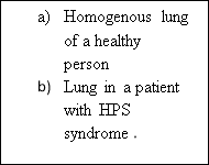
Figure 4: Ventilation-perfusion mismatches the oxygen reach the capillaries and blood.
- The nonuniform perfusion blood flow may lead to the formation of functional intrapulmonary vascular dilations which is the major cause of hypoxemia and the defining feature of HPS [9].
- The hyperdynamic circulation as well as the increased cardiac output which associated with liver disease reduces the transit time of blood in the lung vasculature, thus the time available for the oxygen diffusion is reduced. This in turn contributes to the hypoxemia [9].
- HPS also caused a decreased arterial partial pressure of oxygen (PO2) as a result of the inability of oxygen molecules to diffuse to the center of the dilated pulmonary capillaries to oxygenate the haemoglobin in the erythrocytes [8].


Figure 5 shows illustration of precapillary pulmonary vascular dilatations.
- On the other hand, the increasing of alveolar PO2 with supplement oxygen may increase the blood arterial PO2 and improve the hypoxemia [8].
- HPS patients have been reported to have decreased pulmonary vascular resistance and decreased hypoxic pulmonary vascular constriction [8].
Hepatorenal syndrome (HRS)
Hepatorenal syndrome (HRS) is generally occurs in patients with cirrhosis and portal hypertension [10]. HRS is characterised by major disturbances in circulatory function and renal failure [10]. Intense vasoconstriction of the renal circulation has resulted in renal failure [10].
The HRS is the final consequence of extreme underfilling of the arterial circulation secondary to arterial vasodilatation in the splanchnic vascular bed [10, 11]. The principle abnormality in the systemic circulation is low arterial pressure caused by greatly reduced total systemic vascular resistance [11]. The prognosis remains poor, particularly when there is rapidly progressive renal failure [11].
HRS occurs in the setting of cirrhosis predominantly, but it can also be developed in other types of chronic liver disease like alcoholic hepatitis.

Figure 6: Proposed pathogenesis of HRS in cirrhosis, according to the arterial vasodilatation [13]
- The mechanism of HRS is vasoconstriction of the renal circulation is multifactorial, involving disturbance in the activity and circulatory function of systemic and renal vasoactive mechanisms [13].
- There is severe arterial underfilling in the systemic circulation which is related to the portal hypertension [13]. The arterial underfilling is caused by the vasodilatation of the splanchnic circulation related to increased splanchnic production of vasodilator substances, particularly nitric oxide [17].
- The increased activity of the vasoconstrictor systems caused greatly reduce in renal perfusion and GFR however tubular function is preserved [13,14,16].
- The vasoconstrictor system has resulted in the retention in free water (arginine vasopressin) and retention in sodium (renin-angiotensin and sympathetic nervous system) that occurs in advanced cirrhosis [15, 16].
- In the early phases of decompensated cirrhosis, increased synthesis of renal vasodilator factors (mainly prostaglandins) caused maintenance of renal perfusion within the normal range because of [17].
- In later phases of the disorder, renal perfusion cannot be maintained because maximum activation of vasoconstrictor systems caused by the extreme arterial underfilling, has decreased production of renal vasodilator factors, or both, and lead to HRS developement [17].
- The activation of vasoconstrictor systems also results in vasoconstriction of some vascular beds other than the kidneys, including the legs, arms and brain. Due to the greatly increased local production of vasodilators, the splanchnic area escapes the effect of vasconstrictors [18].
References
- Webmd.com. (2014).Liver failure causes, symptoms, treatments, tests & more. [online] Retrieved from: http://www.webmd.com/digestive-disorders/digestive-diseases-liver-failure [Accessed: 11 Jan 2014].
- Vitaltherapies.com. (2014).Liver disease | vital therapies. [online] Retrieved from: http://vitaltherapies.com/liver-disease/ [Accessed: 11 Jan 2014].
- Hn632campus.wikispaces.com. 2014.HN632campus – Cirrhosis. [online] Available at: https://hn632campus.wikispaces.com/Cirrhosis [Accessed: 11 Jan 2014].
- Buob, S., Johnston, A. N., & Webster, C. R. L. (2011). Portal hypertension: pathophysiology, diagnosis, and treatment.Journal of Veterinary Internal Medicine,25(2), 169-186.
- Livermd.org. (2014).Hepatitis & cirrhosis. [online] Retrieved from: http://livermd.org/hepatitis.html [Accessed: 11 Jan 2014].
- Krowka, M. J., & Cortese, D. A. (1994). Hepatopulmonary syndrome. Current concepts in diagnostic and therapeutic considerations.CHEST Journal,105(5), 1528-1537.
- Fritz, J. S., Fallon, M. B., & Kawut, S. M. (2013). Pulmonary Vascular Complications of Liver Disease.American journal of respiratory and critical care medicine,187(2), 133-143
- Hoeper, M. M., Krowka, M. J., & Strassburg, C. P. (2004). Portopulmonary hypertension and hepatopulmonary syndrome.The Lancet,363(9419), 1461-1468
- Zhang, J., & Fallon, M. B. (2012). Hepatopulmonary syndrome: update on pathogenesis and clinical features.Nature Reviews Gastroenterology and Hepatology,9(9), 539-549.
- Ncbi.nlm.nih.gov. (2014).Hepatorenal syndrome – national library of medicine – pubmed health. [online] Retrieved from: http://www.ncbi.nlm.nih.gov/pubmedhealth/PMH0001519/ [Accessed: 12 Jan 2014].
- Emedicine.medscape.com. (2014).Medscape: medscape access. [online] Retrieved from: http://emedicine.medscape.com/article/178208-overview#a0104 [Accessed: 11 Jan 2014].
- Ginès, P., Guevara, M., Arroyo, V., & Rodés, J. (2003). Hepatorenal syndrome.The Lancet,362(9398), 1819-1827.
- Arroyo, V., Ginès, P., Gerbes, A. L., Dudley, F. J., Gentilini, P., Laffi, G., … & Schölmerich, J. (1996). Definition and diagnostic criteria of refractory ascites and hepatorenal syndrome in cirrhosis.Hepatology,23(1), 164-176.
- Arroyo, V., Guevara, M., & Ginès, P. (2002). Hepatorenal syndrome in cirrhosis: pathogenesis and treatment.Gastroenterology,122(6), 1658-1676.
- Schrier, R. W., Arroyo, V., Bernardi, M., Epstein, M., Henriksen, J. H., & Rodés, J. (1988). Peripheral arterial vasodilation hypothesis: a proposal for the initiation of renal sodium and water retention in cirrhosis.Hepatology,8(5), 1151-1157.
- Schrier, R. W., Niederberger, M., Weigert, A., & Ginès, P. (1994). Peripheral arterial vasodilatation: determinant of functional spectrum of cirrhosis. InSeminars in liver disease(Vol. 14, No. 1, pp. 14-22).
- Martin, P. Y., Ginès, P., & Schrier, R. W. (1998). Nitric oxide as a mediator of hemodynamic abnormalities and sodium and water retention in cirrhosis.New England Journal of Medicine,339(8), 533-541.
- Angeli, P., & Merkel, C. (2008). Pathogenesis and management of hepatorenal syndrome in patients with cirrhosis.Journal of hepatology,48, S93-S103.
Cite This Work
To export a reference to this article please select a referencing stye below:
Related Services
View all


DMCA / Removal Request
If you are the original writer of this essay and no longer wish to have your work published on UKEssays.com then please click the following link to email our support team:
Request essay removal